This is not likely the first, but this account of an observation in 1964 (after Paulings Nobel Prize talk) it does address the dodecahedral shape that Pauling is showing.
From Besson et al. (2020) The Adenovirus Dodecahedron: Beyond the Platonic Story Viruses. 2020 Jul; 12(7): 718.
The adenovirus dodecahedrons were first discovered in Sweden in 1964. While purifying soluble antigens produced during an HAdV3 infection of human cells, Erling Norrby observed a homogenous population of particles sedimenting by centrifugation at a rate corresponding to 50–60 S. They appeared as six- or five-pointed ‘stars’ composed of identical tubular, capsomer-like structures, each associated with a thin club-shaped projection 21. The overall diameter between the points surrounding the star was assessed between 40 and 50 nm. These points were later attributed to the fiber’s knobs, and the tubular capsomer was ascribed to the penton base forming the core (star) of the dodecahedron. These particles do not contain hexons, but are exclusively made of penton base and fiber proteins.
21Norrby E. The relationship between the soluble antigens and the virion of adenovirus type 3. I. Morphological characteristics. Virology. 1966;28:236–248. doi: 10.1016/0042-6822(66)90148-6.
and later:
The adenovirus dodecahedron was ‘rediscovered’ in 1997 by co-expressing the penton base and the fiber of HAdV3 in insect cells using the baculovirus expression system. Insect cells lysates were then subjected to a sucrose gradient and the heavy fractions were analyzed by negative stain electron microscopy, highlighting spherical particles arranged into regular dodecahedrons by their bases31. These particles display two-fold, three-fold, and five-fold symmetry axes. It is worth noticing that, as expected, the HAdV-2 penton base expressed in a similar way did not form dodecahedrons but only pentameric penton bases, as HAdV-2 does not form dodecahedrons under ‘natural’ conditions. This confirms that not all adenovirus serotypes are able to generate such particles. The recombinant dodecahedron made of 12 pentons (12 penton bases with 12 protruding trimeric fibers) was called the Penton Dodecahedron (Pt-Dd) (Figure 2b).
31 Fender P., Ruigrok R.W., Gout E., Buffet S., Chroboczek J. Adenovirus dodecahedron, a new vector for human gene transfer. Nat. Biotechnol. 1997;15:52–56. doi: 10.1038/nbt0197-52.
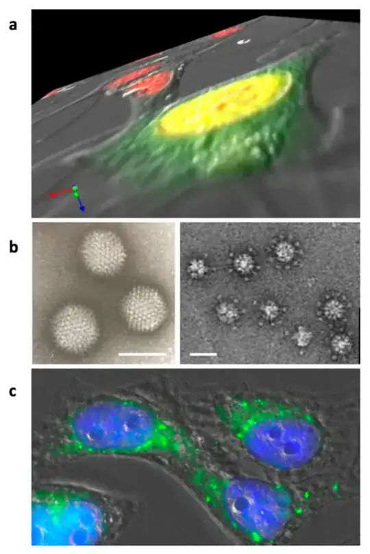
Figure 2. Adenovirus dodecahedron formation and internalization. (a) Z-series of Hela cells infected for 16 h by wt-HAdV3. Nuclei are stained in red and the penton base is detected in green. The penton base is synthesized in the cytoplasm and transported to the nucleus (yellow results from superposition of the red and green signals) where dodecahedron assembly takes place. (b) Electron microscopy images of the purified adenovirus and (Pt-Dd) ‘penton dodecahedrons’ (bars: 90 and 30 nm, respectively). (c) Hela cells incubated with recombinant Pt-Dd for 1 h. Cell shapes were observed by DIC, nuclei are stained in blue, and Pt-Dd are detected in green.

